Everything you need to know about Retinal Detachment
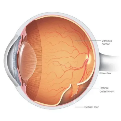
Retinal Detachment refers to a medical emergency in which the light-sensitive tissue, which is located at the back of your eye, gets peeled away from its underlying support. This condition is generally caused by trauma, ageing or even diabetes. Main symptoms which you can notice for this condition include sudden flashes of light, a dark curtain or shadow which seems to spread over your vision and an increase in floaters. In general terms, you may require immediate surgery in order to prevent permanent loss of vision or blindness. You can choose to undergo the surgery in the best eye hospital in Punjab.